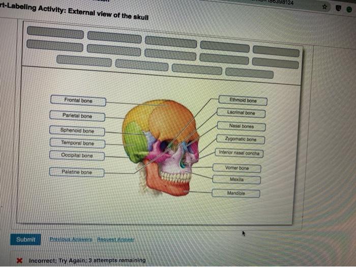
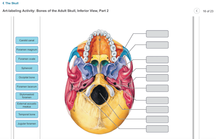
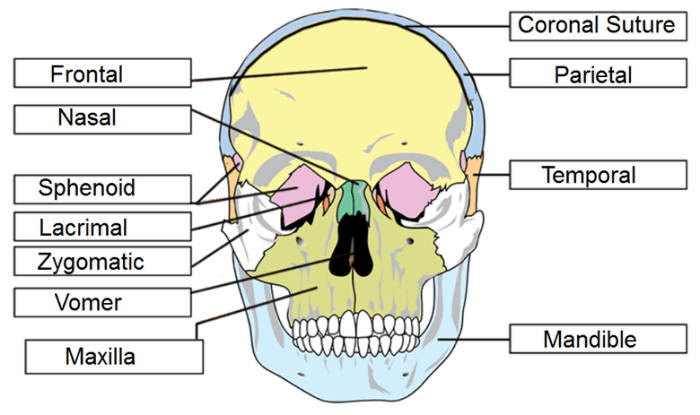
Embarking on an in-depth exploration of the art-labeling activity external view of the skull, this comprehensive guide unveils the intricacies of this anatomical masterpiece. Delving into the realm of osteology, we embark on a journey to decipher the intricate tapestry of the skull’s external features, sutures, foramina, and processes.
Through meticulous labeling and examination, we unravel the secrets of this enigmatic structure, unlocking a deeper understanding of its clinical significance and educational implications.
External Anatomy of the Skull: Basic Overview

The skull, a complex and protective bony structure, serves as the skeletal framework of the head, safeguarding the brain and other vital organs. It comprises a mosaic of flat and curved bones joined by sutures, creating a sturdy yet flexible framework.
The external anatomy of the skull exhibits a myriad of features, including sutures, foramina, and processes, each playing a distinct role in the skull’s overall function.
Bones of the Skull: Identification and Classification
The skull consists of 22 bones, classified into two major groups based on their location and function: the neurocranium and the viscerocranium. The neurocranium, comprising eight bones, forms the protective enclosure for the brain, while the viscerocranium, consisting of 14 bones, forms the facial skeleton, providing support for the eyes, nose, and mouth.
Each bone possesses unique characteristics, allowing for their precise identification.
| Bone Name | Location | Function |
|---|---|---|
| Frontal bone | Forehead | Forms the forehead and protects the frontal lobes of the brain |
| Parietal bones (2) | Sides and top of the skull | Form the sides and roof of the cranium |
| Occipital bone | Back of the skull | Forms the back and base of the cranium, and provides attachment for muscles of the neck |
| Temporal bones (2) | Sides and base of the skull | House the organs of hearing and balance, and provide attachment for muscles of mastication |
| Sphenoid bone | Base of the skull | Forms the middle and lower portions of the skull’s base, and houses the pituitary gland |
| Ethmoid bone | Front of the skull | Forms the roof of the nasal cavity and part of the orbits |
| Maxilla (2) | Upper jaw | Forms the upper jaw, supports the teeth, and houses the maxillary sinuses |
| Mandible | Lower jaw | Forms the lower jaw, supports the teeth, and provides attachment for muscles of mastication |
Landmarks and Regions of the Skull: Detailed Examination
The skull’s external anatomy is characterized by several significant landmarks and regions, each with clinical importance. The frontal bone features the supraorbital margin, which forms the upper border of the orbit, and the glabella, a smooth area between the eyebrows.
The parietal bones display the parietal eminences, which serve as attachment sites for muscles. The temporal bones exhibit the mastoid process, which provides attachment for muscles of the neck, and the styloid process, which provides attachment for muscles of the tongue.
Sutures, Foramina, and Processes: Exploration and Function
Sutures are immovable joints that connect the bones of the skull. The coronal suture joins the frontal bone to the parietal bones, while the sagittal suture joins the two parietal bones. Foramina are openings in the skull that allow for the passage of nerves, blood vessels, and muscles.
The foramen magnum, located at the base of the skull, transmits the spinal cord. Processes are bony projections that provide attachment for muscles or ligaments. The zygomatic process of the temporal bone articulates with the maxilla to form the zygomatic arch, providing support for the cheekbone.
Clinical Applications: Medical Imaging and Surgical Procedures, Art-labeling activity external view of the skull
Art-labeling activities play a crucial role in medical imaging, such as X-rays, CT scans, and MRIs. Accurate labeling of anatomical structures enables precise diagnosis and treatment planning. In surgical procedures involving the skull, accurate labeling ensures safe and effective navigation, minimizing risks to the patient.
Educational Implications: Enhancing Learning and Retention
Art-labeling activities enhance the learning and retention of anatomical structures by engaging multiple senses and promoting active recall. Interactive exercises and games that utilize art-labeling, such as labeling quizzes or 3D skull models, improve student engagement and foster a deeper understanding of the skull’s anatomy.
FAQ Corner: Art-labeling Activity External View Of The Skull
What is the significance of art-labeling activities in medical imaging?
Art-labeling activities play a crucial role in medical imaging, enabling accurate identification and localization of anatomical structures. By precisely labeling X-rays, CT scans, and MRIs, medical professionals can effectively interpret images, diagnose conditions, and plan appropriate interventions.
How does art-labeling enhance learning and retention of anatomical structures?
Art-labeling activities serve as powerful educational tools, promoting active engagement and improving the retention of anatomical information. Through the process of labeling, students actively interact with the material, reinforcing their understanding of complex structures and their relationships.
What are the different types of sutures, foramina, and processes found on the skull?
The skull exhibits a variety of sutures, foramina, and processes, each with distinct functions. Sutures are immovable joints that connect different skull bones, while foramina are openings that allow nerves and blood vessels to pass through. Processes are bony projections or extensions that serve for muscle attachment or articulation with other structures.


